Bioengineered H- Ferritin Nanocages for Quantitative Imaging of Vulnerable Plaques in Atherosclerosi
《动脉粥样硬化》
题目:《Bioengineered H- Ferritin Nanocages for Quantitative Imaging of Vulnerable Plaques in Atherosclerosis》(译:《生物工程化的H-铁蛋白纳米笼用于动脉粥样硬化中易损斑块的定量成像》)
Abstract:Inflammation and calcification concomitantly drive atherosclerotic plaque progression and rupture and are the compelling targets for identifying plaque vulnerability. However, current imaging modalities for vulnerable atherosclerotic plaques are often limited by inadequate specificity and sensitivity. Here, we show that natural H-ferritin nanocages radiolabeled with technetium-99m (99mTc-HFn) can identify and accurately localize macrophage-rich, atherosclerotic plaques in living mice using combined SPECT and CT. Focal 99mTc-HFn uptake was observed in the atherosclerotic plaques with multiple high-risk features of macrophage infiltration, active calcification, positive remodeling, and necrosis on histology and in early active ongoing lesions with intense macrophage infiltration. The uptake of 99mTc-HFn in plaques enabled quantitative measuring of the dynamic changes of inflammation during plaque progression and anti-inflammation treatment. This strategy lays the foundation of using bioengineered endogenous human ferritin nanocages for the identification of vulnerable and early active plaques as well as potential assessment of anti-inflammation therapy.(译:炎症和钙化伴随着驱动动脉粥样硬化斑块的进展和破裂,是识别斑块脆弱性的重要目标。然而,目前针对易损动脉粥样硬化斑块的成像方式通常受到特异性和敏感性不足的限制。在这里,我们展示了用锝-99m ( 99m Tc-HFn)放射性标记的天然 H-铁蛋白纳米笼可以使用 SPECT 和 CT 组合识别并准确定位活小鼠体内富含巨噬细胞的动脉粥样硬化斑块。焦距99m在具有巨噬细胞浸润、活动性钙化、阳性重塑和组织学坏死等多种高危特征的动脉粥样硬化斑块中以及在具有强烈巨噬细胞浸润的早期活动性持续病变中观察到 Tc-HFn 摄取。斑块中99m Tc-HFn的摄取能够定量测量斑块进展和抗炎治疗期间炎症的动态变化。该策略为使用生物工程内源性人铁蛋白纳米笼识别脆弱和早期活动斑块以及抗炎治疗的潜力奠定了基础。)
期刊:ACS Nano
Impact Factor: 13.709
全文链接:点击下载
研究作者:谭辉
研究单位:上海中山医院核医学科
论文中图像及信息:

相关实验信息及图像
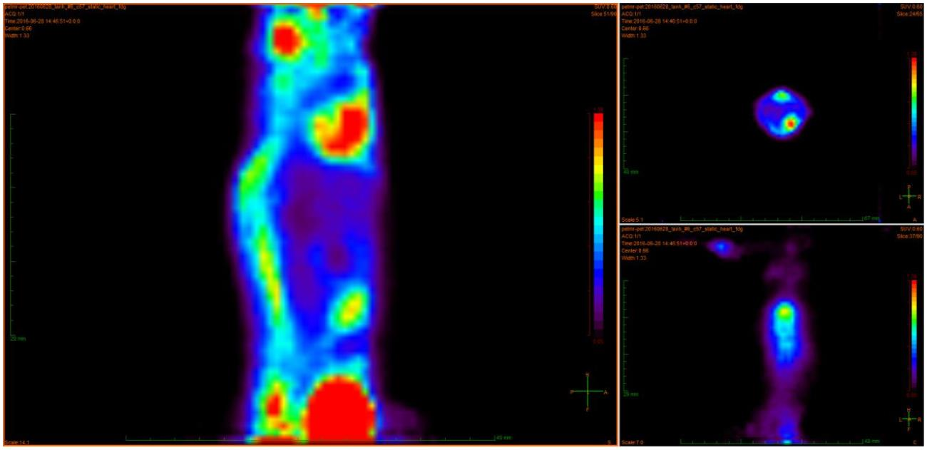
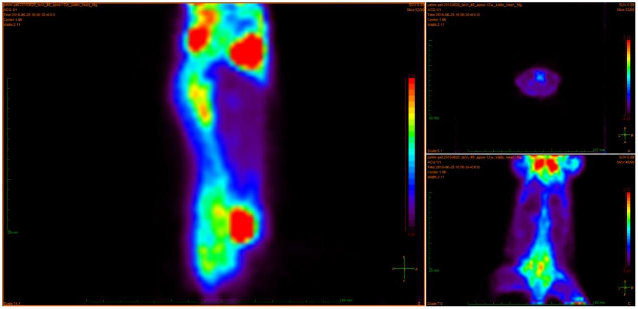
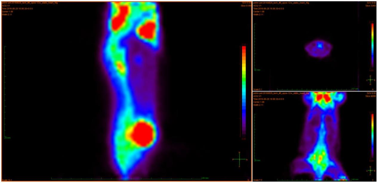
实验一
实验模型:C57BL/6J 小鼠_12 weeks Apoe 模型
显像剂:FDG
给药方式:尾静脉给药
数据采集时间:30min
注射剂量:323uci
研究图像:


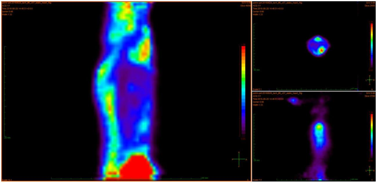
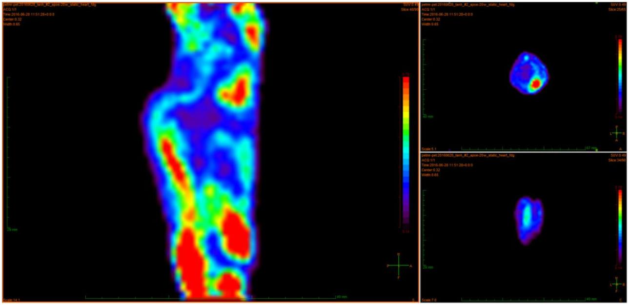
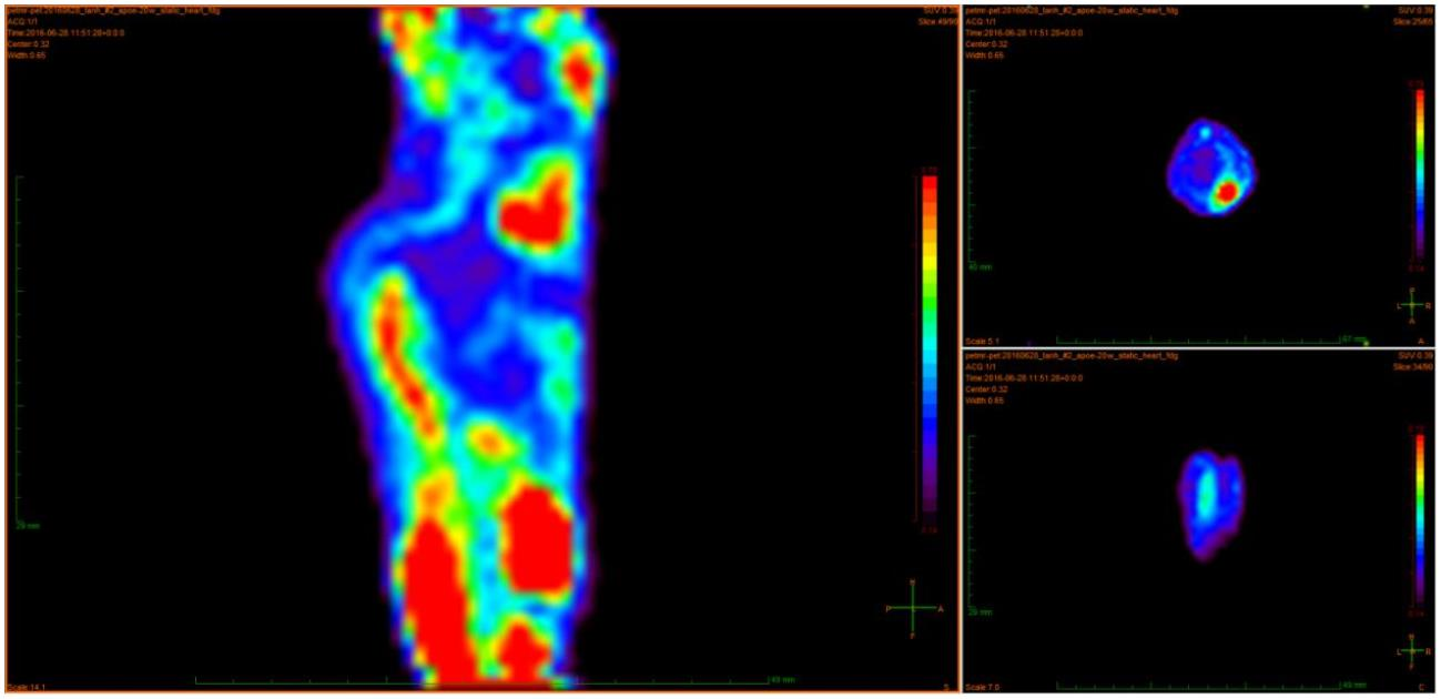
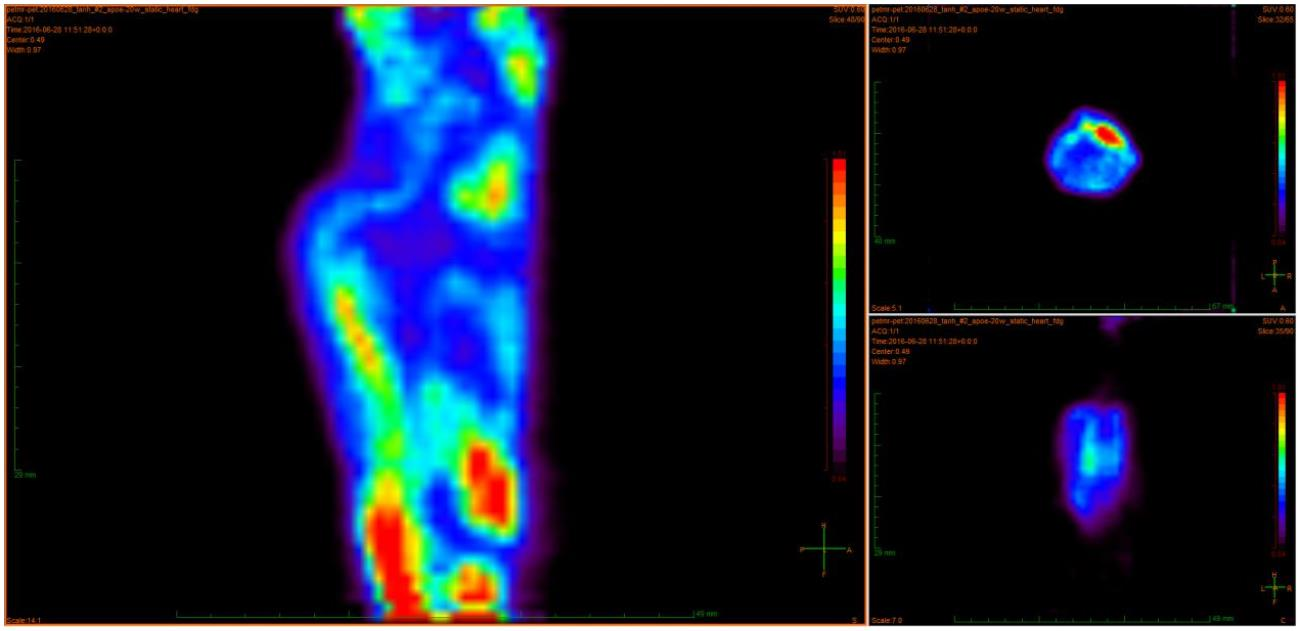
实验二
实验模型:C57BL/6J 小鼠_20 weeks Apoe 模型
显像剂:FDG
给药方式:尾静脉给药
数据采集时间:30min
注射剂量:299uci
研究图像:


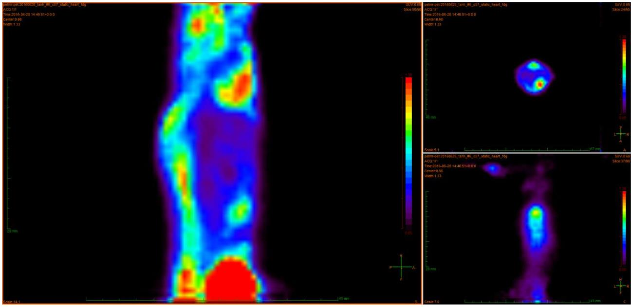
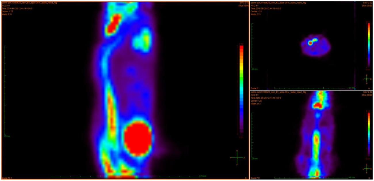
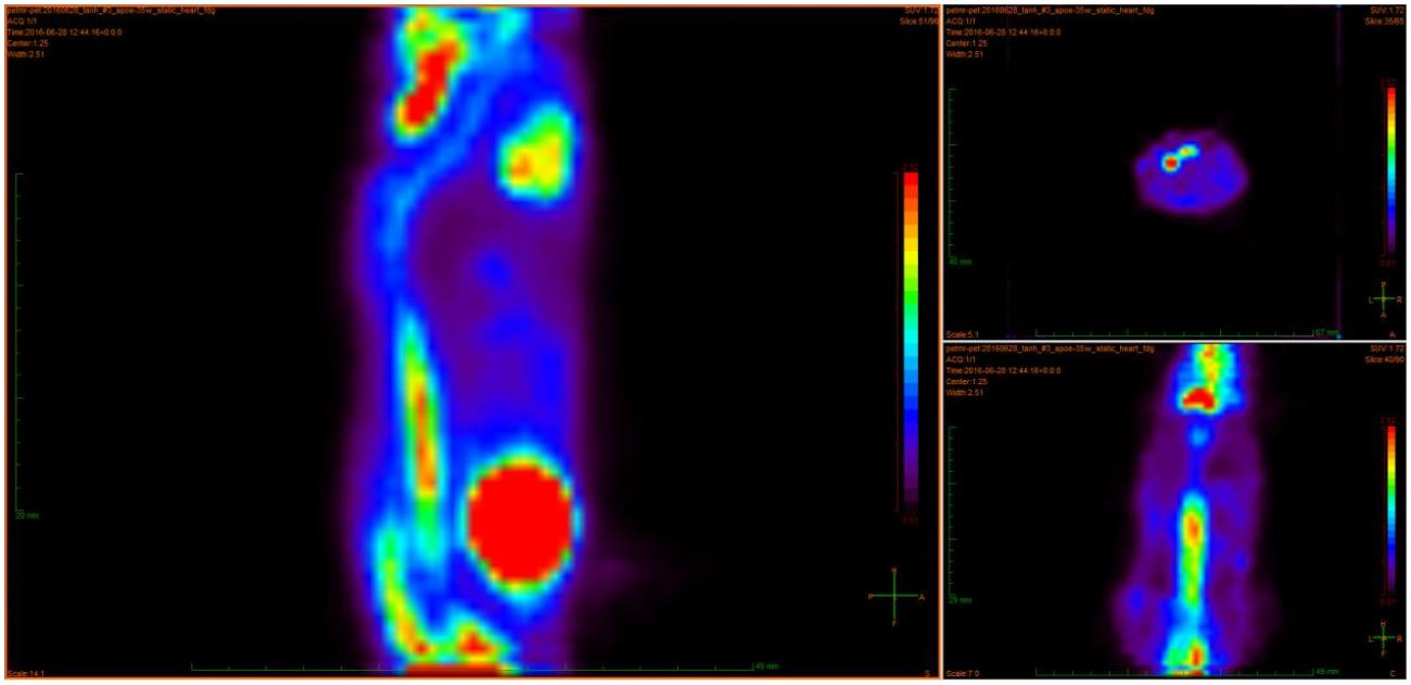
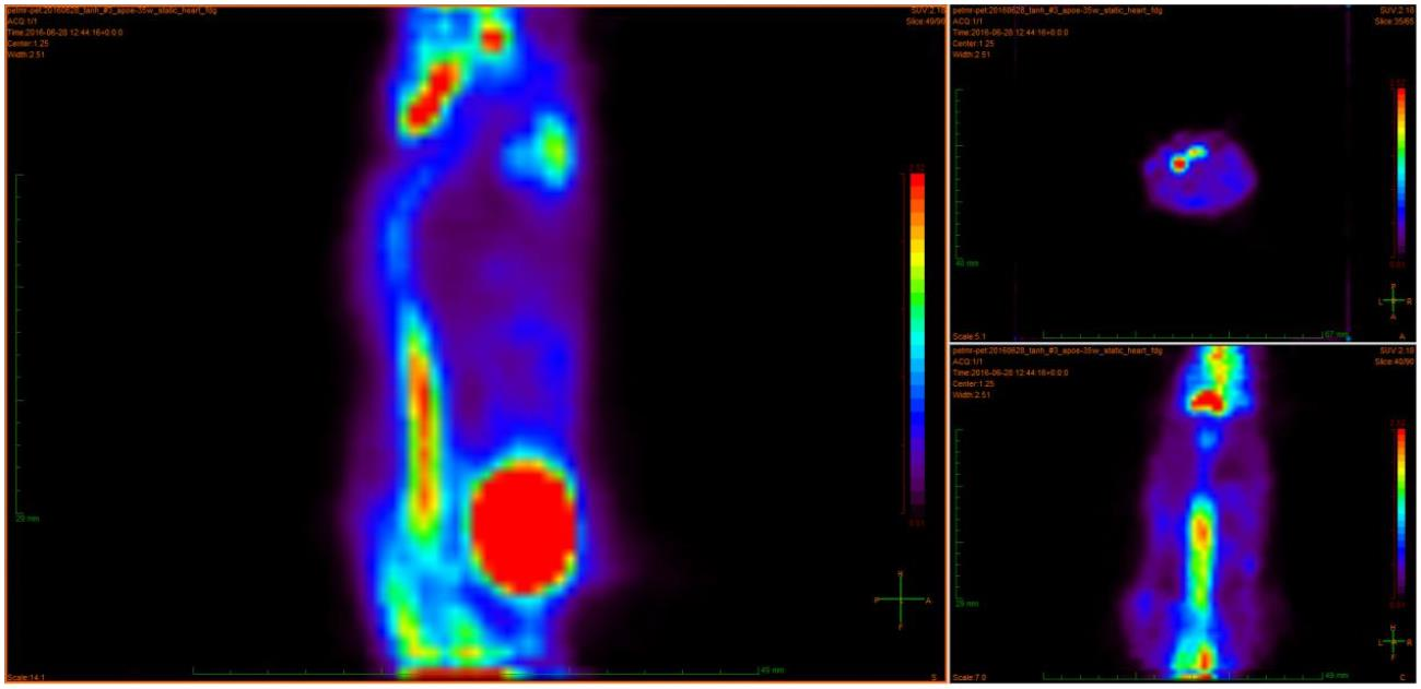
实验三
实验模型:C57BL/6J 小鼠_33 weeks Apoe 模型
显像剂:FDG
给药方式:尾静脉给药
数据采集时间:30min
注射剂量:264uci
研究图像:


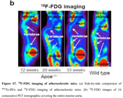
实验四
实验模型:C57BL/6J 小鼠_Wild type Apoe 模型
显像剂:FDG
给药方式:尾静脉给药
数据采集时间:30min
注射剂量:342uci
研究图像: